Mosaic is a digital micromirror device (DMD) used for photo-stimulation applications such as optogenetics, FRAP or photoconversion. It is available in two versions:
Large field of view model: provides a larger working area but lower power densities- suits optogenetics and applications where lower powers are required
Higher power density model: has a smaller working area available for photostimulation but enables higher power densities required for FRAP, photoswitching and conversion applications.
These use the same DMD array, but use different optics to allow for a different power density and illumination area. The Mosaic DMD device may be combined with a range of illumination sources since the Mosaic itself acts as a configurable mirror array. Compatible illumination sources include one, or a combination of: Mosaic Diode lasers, HLE (High Power Laser Engine) and LED illumination.
By using appropriate beam splitters and a suitable microscope cube, imaging and photostimulation wavelengths can be combined. Custom or multiple regions can be defined and the photostimulation illumination delivered precisely and the effects can be observed such as recovery of fluorescence.
Both models provide the following features within Fusion:
-
Triggering
-
Illumination Time
Triggering
Triggering can be configured within the Protocol Manager Tab to that the sample receives photostimulation at the required time period. Refer to the Triggering section for more information.
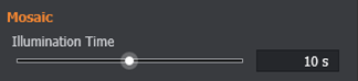
Illumination Time
The illumination time feature changes the duration of photostimulation illumination for the selected region(s). By default it is located in the Channel Manager tab under Channel Settings but can be relocated to Advanced. Mosaic supports illumination durations between 1ms and 200 seconds using the slider or manually entering the required illumination time.
To create photostimulation channels refer to: Setting up and performing Photostimulation with Mosaic
For Calibration of Mosaic see: Mosaic Setup and Calibration
For setting up a protocol see: Creating and Running a Photostimulation Protocol
Suitable Beamsplitter Plugs and Microscopes cubes should be selected to allow photostimulation and imaging wavelengths to reach the specimen see: Beamsplitter Plugs and Microscope Cubes for Photostimulation