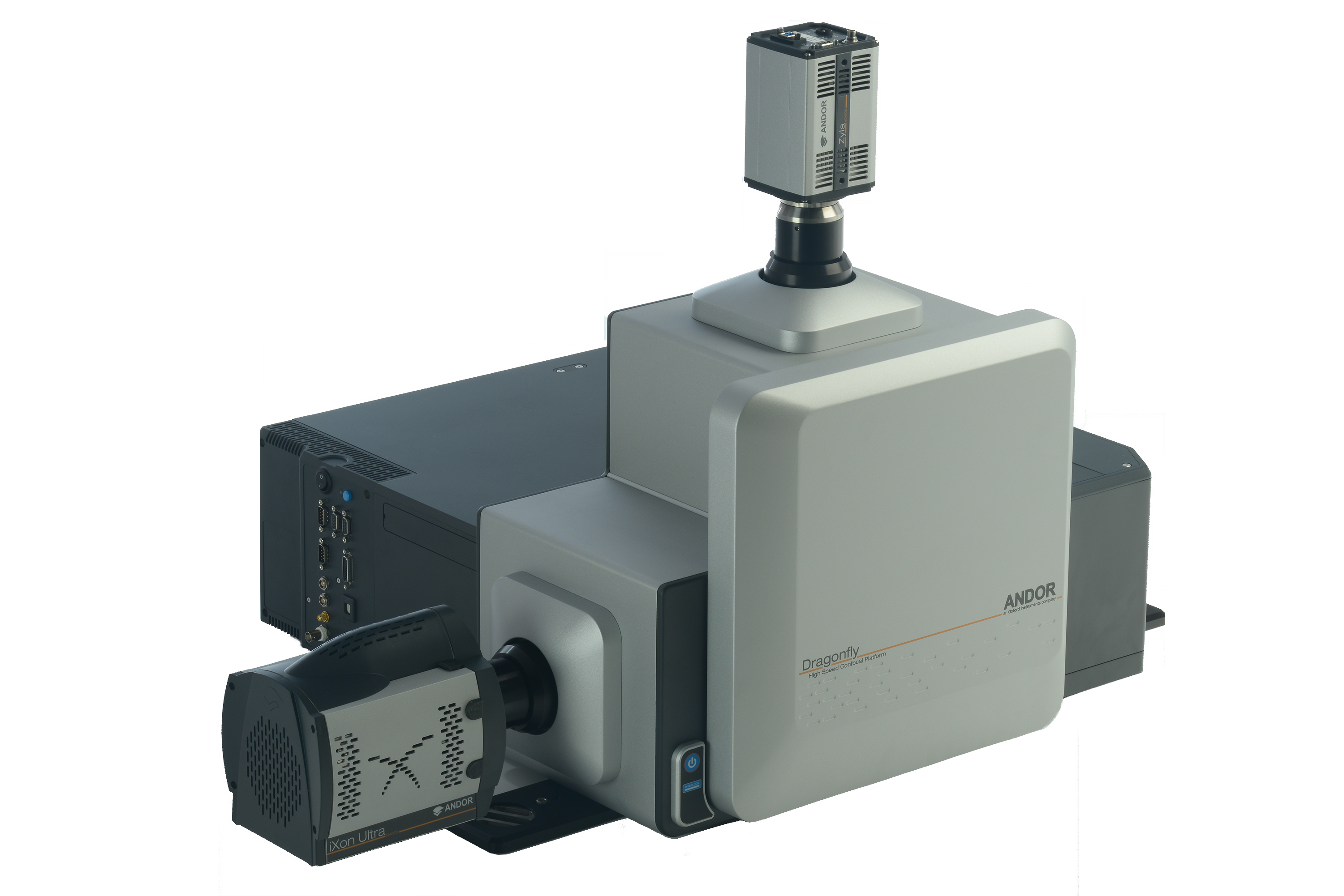
Dragonfly
Introduction
Dragonfly is a multi-modal imaging platform enabling laser-based widefield, spinning-disk confocal, and optionally total internal reflectance fluorescence (TIRF) microscopy when connected to a research-grade microscope. The Dragonfly is intended for professional scientific research applications, especially live-cell biological imaging and photo-stimulation. Fusion has been developed to exploit the full functionality and performance of the Dragonfly.
Overview of the Dragonfly
Laser light from an Integrated Laser Engine (ILE) is fed as an input into one port of the Dragonfly via a multi-mode optical fibre for widefield and confocal illumination (passing first through a Borealis Beam Conditioning Unit – BCU) and/or a single-mode optical fibre through a different port for TIRF illumination.
The Dragonfly couples to an inverted microscope through its left side port using a microscope-specific connector flange and mating optics. Proper and secure mating of the Dragonfly to a microscope requires that the microscope be raised to a certain height using the supplied pedestal kit and fastened onto an optical table benchtop. For laser safety reasons, the Dragonfly should only be aligned and interlocked to the microscope by a qualified Andor representative.
Illumination laser light transferred through the Dragonfly enters the microscope’s left side port and subsequent fluorescence emission light generated by the sample specimen and collected by the microscope optics returns to the Dragonfly through the same port. The emission light is separated from the excitation laser light by a multiband excitation dichroic optic and forms an image on the camera after passing through an appropriate interference filter loaded in a motorized filter wheel.
To find out more about the Dragonfly, please visit the Microscopy section of the Andor website
For further information on using the Dragonfly, please refer to the Dragonfly Hardware Guide, supplied with your system, and Dragonfly Features in this user guide.